Painless Jaundice From a Tiny Tumour: Ampullary Cancer
Yellow eyes and no pain at all. That combination gets taken less seriously than almost anything else in medicine. Pain is what makes people move. Without it, jaundice reads like a liver thing, a virus, something that will pass. It is often the opposite. A painless block in the bile outflow is the presentation most likely to turn out to be a tumour, and the tumour doing it can be smaller than a fingernail. The way through is not a bigger scan. It is refusing to let the first comfortable explanation close the question, and Healz was built to keep asking.
This post walks the finding: where the ampulla of Vater sits and why a tumour there announces itself early, what the bloodwork shows, how the workup runs from imaging to the endoscope, what a biopsy can and cannot settle, and why this cancer is operable more often than its neighbours.
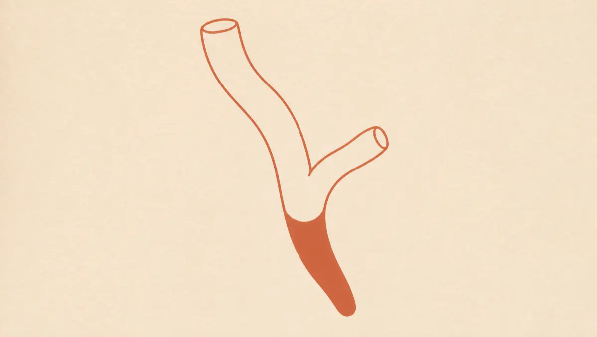
Where the ampulla sits, and why a few millimetres matter
The ampulla of Vater is a junction, not an organ. A 2023 review in Cancers describes the ampullary region as the crossroad of three different structures: the bile duct, the main pancreatic duct, and the duodenal mucosa. Bile made in the liver runs down the common bile duct, meets the pancreatic duct inside the duodenal wall, and empties into the gut through a small raised opening called the major duodenal papilla. Everything the liver sends the intestine passes through that one doorway.
That geometry is the whole story. A tumour in the body of the pancreas can reach several centimetres before it presses on anything that complains. A tumour at the ampulla is sitting in the doorway. A few millimetres of growth closes it, and once it closes, bile has nowhere to go. The cancer becomes visible in a bathroom mirror long before it becomes large. It is also rare: the same review puts ampullary carcinomas at under 1% of gastrointestinal cancers, roughly six cases per million.
Ask Healz.
One chat instead of ten apps. 1M+ rare cases checked. The root cause, not the label. Private.
Why yellow eyes without pain is the version that matters
Jaundice is bilirubin building up in the blood because bile is backing up in the liver, per MedlinePlus. What differs is the character of the block. A stone is intermittent and it hurts, because a stone moves, grinds and inflames. A growing mass is quiet and steady; it just keeps narrowing.
That difference is measured. In a 2008 Journal of Hospital Medicine cohort of patients admitted with obstructive jaundice, abdominal pain at presentation was reported by 34% of those whose obstruction proved malignant, against 71% of those whose obstruction was benign. Pain is not a rule in either direction, and the same study found that 9% of its gallstone patients had painless jaundice. But the absence of pain shifts the odds toward a mass, the exact reverse of how it feels to the person noticing their eyes.
The companion signs are easy to dismiss one at a time. MedlinePlus lists dark urine, clay-coloured or pale stools, itching, nausea and vomiting among the symptoms of bile duct blockage, and itching in particular gets handled as a skin complaint. The whole set arriving together, with no pain anywhere in it, is the pattern that prompts an urgent workup.
What the bloodwork shows when bile cannot leave
The blood says the plumbing is blocked before any scan names the blocker. MedlinePlus lists increased serum bilirubin, increased alkaline phosphatase, increased GGT and increased liver enzymes as results that could be due to a bile duct blockage.
What matters is the shape of that set, not any one value. When alkaline phosphatase and GGT climb out of proportion to the transaminases, the pattern points at the drainage rather than at damaged liver cells; a hepatitis picture runs the other way around. That shape is what an ai lab report reader is for, rather than one flagged value.
Direction carries weight too. A bilirubin that rose, fell and rose again fits something that moves, like a stone, while one that has climbed steadily across three draws fits something that is growing.
How the workup runs, from imaging to the endoscope
MedlinePlus lists the tests used when a bile duct blockage is suspected: abdominal ultrasound, abdominal CT scan, MRCP, ERCP, percutaneous transhepatic cholangiogram and endoscopic ultrasound. In practice these are a sequence, not alternatives. Ultrasound answers one question fast: are the ducts dilated. CT maps the region and looks for spread, which is where the hedged language of a radiology report starts mattering. MRCP photographs the ducts without instrumenting them.
Then the endoscope, where the ampulla has an advantage no other periampullary site has: it can be seen. A scope reaches the papilla directly, so the lesion is looked at and sampled, and ERCP can both image the ducts and relieve the obstruction with a stent in the same session. Endoscopic ultrasound judges how deep the tumour goes and whether nearby nodes are involved, although it is less decisive than it sounds: a meta-analysis of endoscopic ultrasound staging in ampullary tumours found pooled sensitivity of 77% and specificity of 78% for identifying T1 lesions, and 70% and 74% for detecting nodal involvement.
The same picture of painless obstruction can come from the bile duct itself, which our guide to cholangiocarcinoma and bile duct cancer covers, or from a lesion in the head of the pancreas. Sorting out which of the three is much of what the imaging and endoscopy exist for.
What a biopsy result can and cannot settle
A biopsy at the papilla is good at finding an adenoma and much weaker at finding the cancer growing inside one. A review in the World Journal of Methodology reports forceps biopsy sensitivity above 90% for demonstrating an adenoma but a miss rate for carcinoma of up to 30%, and states plainly that a negative diagnosis of carcinoma on biopsy of an ampullary adenoma does not exclude a focus of adenocarcinoma. The malignant part can sit under a benign surface, and the forceps took the surface.
So the exact wording on the pathology report is load-bearing. "No evidence of malignancy in the fragments submitted" is a different sentence from "no malignancy", and the gap between them is the gap between a follow-up plan and false reassurance. This is where an AI second opinion earns its keep.
Two subtypes under one name, and the operation that can cure it
Under the microscope, ampullary cancers sort mainly into two lineages: intestinal, resembling the duodenal lining beside it, and pancreatobiliary, resembling duct tissue. In a series of 170 resected patients published in Scientific Reports in 2019, subtyping was possible in 119: pancreatobiliary in 58%, intestinal in 34.5%, mixed in 7.6%. Five-year survival was 27.5% for the pancreatobiliary subtype against 61% for the intestinal one.
That gap is real but it is not settled science. The 2023 Cancers review notes that in the ESPAC-3 trial population there was no difference in overall survival between the subtypes, and that in one analysis subtype was not associated with survival once other factors were accounted for. So the subtype is a strong signal across many series rather than a proven independent predictor.
The curative operation is a pancreaticoduodenectomy, the Whipple, which removes the head of the pancreas, the duodenum, the gallbladder and the bile duct and rebuilds the connections. It is serious surgery: the World Journal of Methodology review reports morbidity of roughly 25% to 65% and mortality of 0% to 10%, under 4% at high-volume centres, and notes it is more likely to achieve complete excision than local removal, which is reserved for adenomas and very early lesions.
The anatomy pays off at this stage. The 2023 Cancers review reports that over 80% of ampullary carcinomas are resectable, with five-year survival rates reaching over 30% to 50%, significantly higher than in cholangiocarcinoma and pancreatic cancer. That is not because the cells are kinder: a tumour in the bile doorway forces the issue while it is small, whereas a pancreatic tumour a centimetre away can stay silent for months. What follows surgery is less certain: the same review states there is a lack of strong evidence guiding systemic or locoregional therapy, and NCCN guidance includes observation after resection. A lesion found nearby on an incidental scan raises its own questions, which our guide to telling a pancreatic cyst from cancer works through.
How Healz reads a painless jaundice case
A bilirubin trend, a CT report and a paragraph of biopsy wording are one case. They arrive as three, weeks apart, in a lab portal, a scan letter and a pathology PDF. Healz keeps them in one chat, not ten apps, and Frontier AI works the whole file, not the page in front of you.
Healz is equipped with root-cause technology, so a painless jaundice is treated as a question about the outflow instead of a label. It asks what a rushed visit skips, cross-checks the case against more than a million rare cases, and does not stop at "no malignancy in the fragments submitted" when that sentence is the known weak point of the test. Healz's memory pulls the medical facts out of every upload, keeps abnormal values up front and dates each finding, so this week's bilirubin is read next to the one from six weeks ago and a steady climb reads as a climb.
A panel, a CT report and a pathology report uploaded to one chat are read together, which is what a search for a pathology report ai or ai ct scan analysis is really after. For a rare cancer, that is the ai oncology use case that matters: not a label, a worked case. You can bring a board-certified doctor into the same chat for a second opinion.
Frequently asked questions
- Does painless jaundice mean cancer?
Not by itself, but it raises suspicion. In a 2008 Journal of Hospital Medicine cohort of patients with obstructive jaundice, pain was present in 34% of malignant cases and 71% of benign ones, so painlessness tilts the odds toward a mass rather than a stone. It is not a rule in either direction, which is why the pattern is worked up rather than observed.
- Can ampullary cancer be missed on a biopsy?
Yes. A review in the World Journal of Methodology reports that forceps biopsy at the papilla detects adenoma with sensitivity above 90% but misses carcinoma in up to 30% of cases, and that a negative result does not exclude a focus of adenocarcinoma within an adenoma. Sampled fragments are not the whole lesion.
- Is ampullary cancer the same as pancreatic cancer?
No. They are neighbours in the same few centimetres and share the same operation, but they start in different tissue. The 2023 Cancers review reports over 80% of ampullary carcinomas are resectable, far more than in pancreatic cancer, largely because the ampullary tumour declares itself through jaundice while still small.
- What does the intestinal or pancreatobiliary subtype change?
It changes the expected outlook, not the surgery. A 2019 Scientific Reports series found five-year survival of 61% for the intestinal subtype against 27.5% for the pancreatobiliary subtype, though the ESPAC-3 population showed no difference, so it reads as one strong signal among several.
Painless jaundice is the quietest serious finding in the abdomen, and the quiet is the whole problem, which is why Healz keeps asking why the bile stopped instead of accepting the first calm answer.
Written by Healz Team · Filed under Health Insights