A Pineal Region Mass in a Teenager: Germinoma and Its Markers
The complaints arrive in the wrong order. Double vision in a sixteen-year-old often gets an optician's appointment. Headaches get blamed on screens and late nights. Then somebody notices that the eyes will not look up, and the scan that follows shows a small mass in the middle of the brain, where a dozen different tumours sit and look alike on the same picture. Working out which of them it is, before anyone operates, is what this post walks through.
Why a mass this small backs up the whole brain
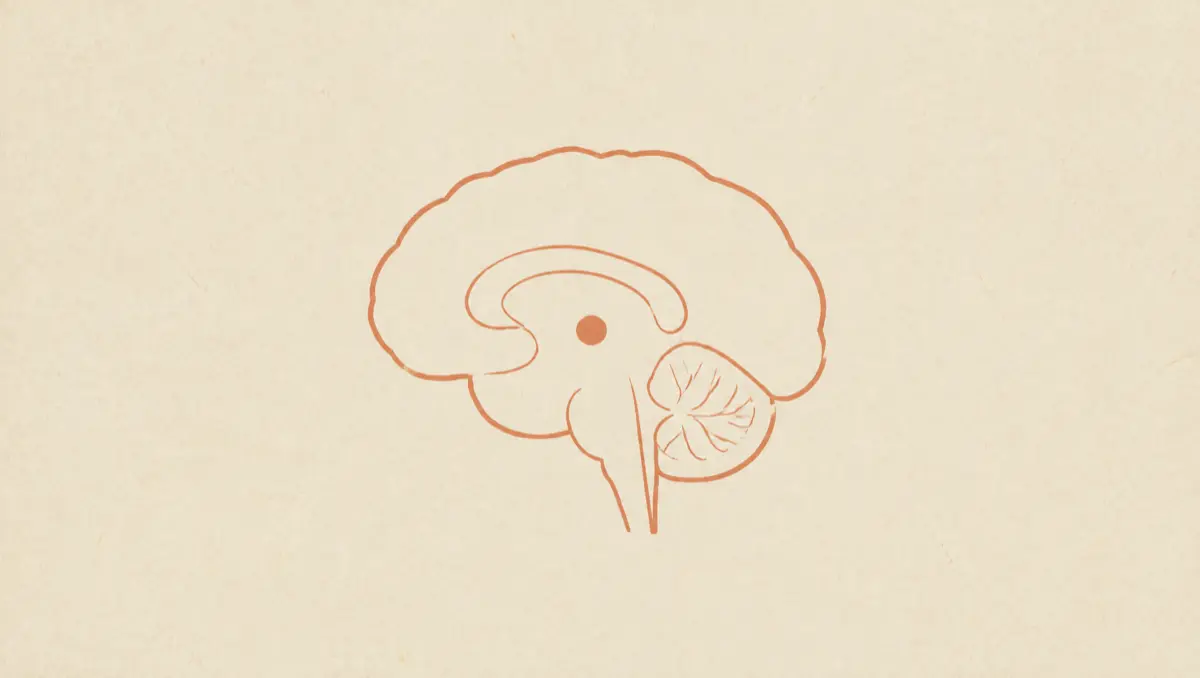
The pineal gland is attached to the back of the roof of the third ventricle, right beside the narrow channel that drains fluid out of it. A germinoma growing forward from there can block that channel, which is how the pressure builds (Eye and Brain, 2023).
Hydrocephalus in pineal region tumours is usually caused by the tumour obstructing the cerebral aqueduct, per a 2023 review of pineal germinoma in the journal Eye and Brain, which puts it at perhaps 90% of patients at presentation and cites a series in which 43 of 56 patients, 81%, had it confirmed radiographically. Papilloedema, the swelling at the back of the eye that raised pressure produces, is found in about 60% of pineal region tumours.
Ask Healz.
One chat instead of ten apps. 1M+ rare cases checked. The root cause, not the label. Private.
The NCI's PDQ summary on childhood central nervous system germ cell tumours notes that patients with tumours in the pineal region usually have a shorter history of symptoms than patients with tumours of the suprasellar or basal ganglionic region, with raised intracranial pressure and diplopia related to tectal and aqueductal compression. Our piece on the headache patterns that get a brain scan covers how a pressure headache separates itself from an ordinary one.
What eyes that will not look up are telling the team
The eye findings localise the lesion before any imaging, because the structures that control vertical gaze sit in the dorsal midbrain, immediately under the pineal gland.
Grouped together they are called Parinaud syndrome, and the Eye and Brain review reports it in up to 75% of patients. The most characteristic sign is a supranuclear palsy of conjugate upward gaze, present in 69.7% of patients, with downward gaze usually spared. Alongside it: semi-dilated pupils that respond poorly to light but still constrict for near vision, called light-near dissociation; convergence-retraction nystagmus, where trying to look up pulls the eyes inward and back; and upper eyelid retraction, known as Collier's sign. Diplopia occurs in 74.3% of patients. The NCI lists Parinaud syndrome, meaning vertical gaze impairment, convergence nystagmus and light-near pupillary response dissociation, along with headache and nausea and vomiting, among the signs unique to masses in the pineal and posterior third ventricular region. Collier's sign comes from the Eye and Brain review, not that list.
A visual field deficit signals something else: the Eye and Brain review notes it suggests a bifocal tumour with suprasellar extension onto the optic chiasm. Some patients present with lesions in both the suprasellar and pineal regions, and the NCI notes the second lesion is often asymptomatic and found on MRI. In suprasellar disease, diabetes insipidus from antidiuretic hormone deficiency occurs in 70% to 90% of patients and is the most common sentinel symptom, per the NCI; the Eye and Brain review puts it at up to 41% of pineal germinomas and 93% of suprasellar germinomas.
A dozen different tumours can sit there
The pineal region is the commonest site of origin for childhood central nervous system germ cell tumours, at 45%, with the suprasellar region second at 30%, per the NCI. In Western countries these tumours peak between the ages of 10 and 19, and male patients have a preponderance of pineal region primaries.
What the scan cannot settle is which tumour it is. The germ cell family splits into germinoma and the non-germinomatous germ cell tumours, which the NCI lists as embryonal carcinoma, yolk sac tumour, choriocarcinoma, teratoma whether mature or immature, teratoma with somatic-type malignancy, and mixed germ cell tumour. Outside that family the same location holds pineoblastoma and pineocytoma, which arise from the pineal gland's own cells, tectal gliomas, and central nervous system lymphoma. The Eye and Brain review says CT and MRI have an 85% to 90% predictive value for germ cell tumours but cannot completely separate central nervous system germinomas and lymphomas from other tumours, which is why imaging alone usually is not the end of the workup.
Benign pineal cysts, which are not tumours, are common: a 2007 study of 100 healthy young volunteers on high-resolution MRI found pineal cysts in 23%, mean largest diameter 4.3 millimetres (range 2 to 14), a rate the authors said matched earlier autopsy studies; the two subjects with long-term follow-up showed no change (Pu et al., AJNR American Journal of Neuroradiology, 2007). Germ cell tumours arise outside the brain too, and the same markers do similar work there, as our piece on germ cell tumours of the ovary in young women sets out.
The markers that can set the plan before any tissue
AFP, alpha-fetoprotein, and beta-hCG, the beta subunit of human chorionic gonadotropin, are the two most useful markers, and the NCI is specific that they are measured in both serum and cerebrospinal fluid. A marker can be raised in only one of the two compartments, which is why both get drawn.
What they can do is remove the biopsy from the sequence, and the NCI states that route twice with different conditions: a biopsy is often required except when the characteristic imaging features are present and increased markers are found in the serum and/or spinal fluid, and, separately, that if preoperative spinal fluid can be obtained safely and markers are found to be elevated, histological confirmation may not be needed. A clearly raised-marker picture points away from pure germinoma and toward a non-germinomatous tumour, which the NCI treats as a separate disease with its own regimen, and AFP is elevated with yolk sac tumours, per the Eye and Brain review. The direction is the NCI's, and the review supplies the counterweight: even pathologically pure germinomas can carry a beta-hCG above 50 mIU/mL.
What they cannot do is confirm germinoma. The NCI's patient summary says it directly: tumour marker levels are not used to diagnose germinomas. That is not the contradiction it looks like beside the no-biopsy route: raised markers can identify a non-germinomatous tumour without tissue, but no marker value proves a germinoma. Germinomas can secrete small amounts of beta-hCG, usually below 50 mIU/mL, the same quantity as 50 IU/L, and the Eye and Brain review notes no strict universal cutoff has been established. The NCI says the levels used to separate germinoma from the non-germinomatous tumours, 50 IU/L in Europe and 100 IU/L in North America, are questionable. Teratomas usually come with negative markers, which the Eye and Brain review states flatly, though the NCI's own marker table allows AFP to be equivocal in immature teratoma and positive when an immature teratoma carries malignant components.
So when markers are negative, mildly elevated below the diagnostic criteria, or atypical, an endoscopic or open biopsy is needed to make a definitive diagnosis, per the NCI.
Why the staging reads the whole spine
These tumours seed the spinal fluid. In addition to whole-brain MRI, MRI of the spine is required, per the NCI, and positive spinal fluid cytology or drop metastases, meaning spinal or cranial subarachnoid deposits, count as metastatic disease. The Eye and Brain review reports drop metastases in up to 30% of pineal tumours and 11% of germinomas at presentation.
The Eye and Brain review prefers the spinal MRI before the lumbar puncture, because the puncture itself can produce artefactual dural enhancement that reads as disease, though the same review notes Mark et al. found that enhancement in only 3.9% of patients imaged after a puncture. And the NCI is explicit that ventricular spinal fluid does not substitute for staging cytology. Cytology is not dependable for diagnosis either: the Eye and Brain review reports tumour cells in the fluid in only 38% of germinomas.
The stage sets the radiation field. The NCI reports excellent results in metastatic germinoma treated with craniospinal irradiation of 24 Gy plus a local boost, a far wider field than localised disease needs.
One operation for the pressure and the tissue
When a biopsy is needed, one route often solves both problems. A single neuroendoscopic procedure can sample the spinal fluid for markers, perform a third ventriculostomy to relieve the hydrocephalus, and biopsy the tumour, per the Eye and Brain review, which cites Hayashi et al.'s endoscopy series: 96.6% diagnostic accuracy for germ cell tumours, 169 of 175 tumours. The limits are named too: the biopsy can miss a teratomatous component, and ventricular bleeding can obscure the view.
Germinomas are highly chemosensitive and radiosensitive tumours, per the NCI, and that sensitivity gets used deliberately to shrink the radiation: neoadjuvant chemotherapy allows reduced radiation therapy doses and volumes. For localised germinoma the NCI describes radiation covering the whole ventricular system at 24 Gy followed by a boost to the primary site of 30 to 40 Gy, and in the Children's Oncology Group trial ACNS1123 patients with a complete response to chemotherapy received 18 Gy of whole-ventricle irradiation with a 12 Gy boost. Focal radiation alone is not recommended, because the NCI says it results in ventricular relapses. Every gray that comes off the plan comes off a brain with decades of use ahead of it.
How Healz holds the eye findings, the markers and the scan as one case
The eye findings, the serum and spinal fluid marker results and the scan report are three pieces of one case, and they usually live in three departments. Healz holds them in one chat with the dates attached.
Healz is equipped with Frontier AI, so the case is worked whole: the AFP and beta-hCG values read across both compartments, against the date the double vision started, rather than one lab slip at a time. A cancer ai reading the MRI report with the markers beside it is reading a case, not a document.
Healz is equipped with root-cause technology, so it does not stop at the first label. It asks what a rushed appointment skips and cross-checks the case against more than a million rare cases, which is how a marker-negative mass keeps teratoma, pineoblastoma and lymphoma on the list until something takes them off.
Healz's memory pulls the medical facts out of every upload, dates them, keeps abnormal values up front and connects them across months, so the beta-hCG from the first admission sits beside the one drawn after chemotherapy. You can bring a board-certified doctor into the same chat for a second opinion.
Frequently asked questions
- What does a mass in the pineal region in a teenager usually turn out to be?
The pineal region accounts for 45% of childhood central nervous system germ cell tumours, per the NCI. Germinoma is the most common single type, but the same spot also holds non-germinomatous germ cell tumours, pineal parenchymal tumours, tectal glioma, lymphoma and the very common benign pineal cyst.
- Can a brain germinoma be diagnosed without a biopsy?
Sometimes a germ cell tumour is diagnosed without tissue, but not germinoma specifically. The NCI states that if preoperative spinal fluid can be obtained safely and markers are elevated, histological confirmation may not be needed, while its patient summary says marker levels are not used to diagnose germinomas.
- What is Parinaud syndrome?
It is the group of eye findings produced by pressure on the dorsal midbrain: impaired upward gaze, pupils that respond poorly to light but still constrict for near vision, and convergence-retraction nystagmus, per the NCI. The Eye and Brain review reports it in up to 75% of patients with pineal germinoma.
The complaints arrived in the wrong order, and the answer gets assembled from an eye exam, two fluids and one scan read against each other.
Written by Healz Team · Filed under Health Insights