A Mass Behind the Breastbone: Thymoma and Myasthenia Gravis
An eyelid starts to droop by evening. Reading makes the page double. A neurologist orders an antibody test, names myasthenia gravis, and starts treatment. Separately, months earlier or months later, a scan ordered for a cough shows a shadow in the front of the chest. Two clinics. Two charts. Two problems, as far as anyone can see. They are often one disease. Reading the eyelid and the chest shadow as a single finding instead of two unrelated ones is the discipline Healz was built on.
This post walks the pair from the top: where thymic tumours sit, what separates a thymoma from a thymic carcinoma, how often each condition carries the other, what the acetylcholine receptor antibody does and does not answer, why a surgeon may decline to biopsy, and how the tumour is staged and treated.
What the thymus is, and why the address matters
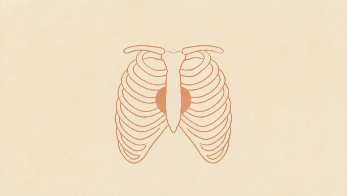
The thymus is a small organ that lies in the upper chest above the heart and under the breastbone, part of the lymph system, making lymphocytes that fight infection (NCI). It does most of that work early in life, is largest around the teenage years, and is gradually replaced by fatty tissue through adulthood, which is part of why a tumour arising there tends to show up in middle age.
The address is the first clue on a chest scan. Radiologists divide the mediastinum, the compartment between the lungs, into front, middle and back. Thymic epithelial tumours, the family that includes both thymoma and thymic carcinoma, are the most common tumours of the anterior mediastinum in adults, although they are uncommon cancers overall (NCI). So the report question is not only how big the mass is but which compartment it sits in.
Ask Healz.
One chat instead of ten apps. 1M+ rare cases checked. The root cause, not the label. Private.
Many of these are quiet. Most people have no symptoms when a thymoma is found, and the warning signs, where they exist, are unglamorous: a cough that will not go, shortness of breath, chest pain, a hoarse voice (NCI). About half are diagnosed while still localized within the thymic capsule (NCI).
Thymoma or thymic carcinoma, and why the distinction runs the plan
They are not two names for one thing. In a thymoma, the cancer cells look a lot like the normal cells of the thymus, grow slowly, and rarely spread beyond the thymus. In a thymic carcinoma, the cells do not look like normal thymus cells, grow more quickly, are more likely to spread, and are harder to treat (NCI). Thymic carcinomas account for approximately 20% of all thymic epithelial tumours, with a greater propensity for capsular invasion, metastases and recurrence (NCI).
Two cautions sit either side of that line. All thymic epithelial tumours have malignant potential and the ability to metastasize, so "benign thymoma" is not an accurate reading of a slow one (NCI). At the same time, behaviour ranges from relatively indolent to aggressive, which is why pathologists sort thymomas into types A, AB, B1, B2 and B3. Those subtypes differ in how often they travel with autoimmune disease.
The imaging offers a hint before any tissue is seen. On contrast CT, smooth round contours have been associated with type A thymoma and irregular contours with thymic carcinoma (NCI). A hint is all it is, which is why radiology wording is hedged and a reader chasing a verdict comes away frustrated.
The eyelid and the chest shadow belong to one disease
Myasthenia gravis happens when the body produces antibodies that block muscle cells from receiving messages from nerves, and double vision and drooping eyelids are the most common first symptoms (MedlinePlus). The weakness worsens with use and eases with rest, and it can reach chewing, swallowing, speech and the chest-wall muscles that drive breathing (MedlinePlus).
The overlap with thymoma is not a curiosity. In reported series, approximately 30% to 65% of patients with thymoma have been diagnosed with myasthenia gravis (NCI), and the rate tracks the subtype: around 71% for type B2 and 57% for B1, against roughly 17% for type A and 16% for AB (NCI). Myasthenia gravis is the most common autoimmune paraneoplastic disease linked with thymoma, and not the only one. Thymoma-associated hypogammaglobulinemia, also called Good syndrome, and pure red cell aplasia are also described, and other autoimmune paraneoplastic conditions can involve any organ (NCI).
Read from the other direction the number is smaller and the published range is wide. Reviews report thymoma in something like 10% to 30% of people with myasthenia gravis, and estimates vary that much because populations and diagnostic criteria differ, with many cohorts recruited from surgical centres. That is the honest version: a minority, and no one tidy figure for it. What follows from it is routine rather than exotic. Chest imaging is part of the standard workup of myasthenia gravis, and MedlinePlus lists a CT or MRI scan of the chest to look for a tumour among the tests used.
What the antibody answers, and what it does not
The confirming blood test in myasthenia gravis checks for acetylcholine receptor antibodies and others associated with the disease (MedlinePlus). In thymoma-associated myasthenia gravis the autoantibodies can hit several neuromuscular targets, particularly the acetylcholine receptor and titin, a striated muscle antigen (NCI).
Here is the part that gets over-read. A positive acetylcholine receptor antibody confirms autoimmune myasthenia gravis. It is not a test for a thymus tumour. In a review of 1,141 patients with myasthenia gravis, the negative predictive value of acetylcholine receptor binding antibodies for thymoma was 99.7% across all ages, so patients without the antibody are unlikely to have one. The positive predictive value ran the other way: highest at 50.0% when those antibodies plus striated muscle antibodies were present and the disease began before age 40, and under 9% for all antibodies after 40. The authors concluded the antibodies are not diagnostic for thymoma, though in early-onset disease their presence should raise suspicion for it (Choi Decroos and colleagues, Muscle and Nerve, 2014).
Two questions, two instruments. The antibody answers whether the weakness is autoimmune myasthenia gravis. The chest imaging answers whether there is a tumour in the thymus. A blood result read as covering both is where the chest shadow goes unlooked-for, a pattern that also runs through how a mediastinal mass is worked up and through paraneoplastic syndromes that announce a hidden cancer.
Why the mass may go straight to the operating room
Patients are often surprised that the plan skips the biopsy. The reason is about not spilling tumour cells.
Intravenous contrast CT is the workhorse for diagnosis and clinical staging, usually accurate for tumour size, location and invasion of vessels, the pericardium and the lungs, while MRI is considered superior for assessing chest wall invasion (NCI). When a mediastinal mass is highly suspicious for an early-stage thymic epithelial tumour and looks completely resectable, surgical resection serves as both the diagnostic and the therapeutic procedure (NCI).
The caution is specific. Surgical biopsy is avoided when thymoma is strongly suspected and resectable, and biopsies that violate the pleural space are avoided because of reported pleural seeding, which can turn an early-stage tumour into a widely staged one (NCCN guidance as summarized in a Journal of Thoracic Disease review, which also notes seeding is reported at exceedingly low rates). When the tumour looks locally advanced and unresectable, tissue is obtained by core needle biopsy or an open biopsy that avoids a transpleural route.
Staging, surgery and what the follow-up is for
Surgery is the main treatment, especially for early-stage disease, with en bloc resection for invasive tumours where that is possible (NCI). Postoperative radiation therapy is associated with a survival benefit and is generally recommended for stage II or stage III disease (NCI). Locally advanced disease is handled with multimodality therapy including chemotherapy and radiation, and systemic therapy alone is used for metastatic tumours (NCI).
Two staging systems appear on reports, which is a frequent source of confusion. The Masaoka system, proposed in 1981 and modified by Koga in 1994, is the most commonly used, and the modified version is recommended by the International Thymic Malignancies Interest Group. The AJCC and UICC have also adopted a TNM classification built from a database of more than 10,000 subjects, applying to both thymoma and thymic carcinoma (NCI). A Masaoka stage and a TNM stage side by side are not two different answers.
When myasthenia gravis is in the picture, the neurology and the surgery get planned as one thing rather than in sequence, because myasthenic weakness can worsen after the operation; postoperative myasthenic crisis is a recognised complication with its own literature, including a 2020 systematic review of risk factors in the Journal of Cardiothoracic Surgery. Follow-up is long: thymomas may recur long after treatment is completed, thymic carcinoma recurs more often, and the risk of a second, different cancer is raised, so follow-up is lifelong (NCI).
How a neurology note and a chest scan get read together
A neurology note and a chest scan mean one thing read together and almost nothing read in sequence, months apart, by two people who never see the other document. Closing that gap is the point of putting the case in one place instead of ten apps and three portals.
Healz is equipped with root-cause technology. That is why it does not stop at the first label: it asks what the eyelid has to do with the shadow, cross-checks the case against more than a million rare cases, and drills toward the cause rather than the symptom in front of it. Frontier AI works the whole file at once, so an ai ct scan analysis of the imaging report sits beside the antibody result and the neurology letter, not in a separate tab. Healz's memory pulls the medical facts out of every upload, keeps abnormal values up front, dates each finding and connects them across months, so a subtype written on a pathology report in spring is still there when the next scan lands. Cancer AI reading your own documents in a private encrypted library is a different proposition from a search box, and an AI second opinion built that way is worth asking for. You can bring a board-certified doctor into the same chat for a second opinion.
Frequently asked questions
- Does a thymoma always cause myasthenia gravis?
No. Across reported series, approximately 30% to 65% of people with a thymoma have been diagnosed with myasthenia gravis, so a substantial share never develop it (NCI). The rate varies by subtype, around 71% for type B2 and roughly 16% to 17% for types A and AB (NCI).
- Can myasthenia gravis be the first sign of a thymus tumour?
It can be the finding that prompts the chest to be imaged. Chest imaging is part of the standard evaluation of myasthenia gravis, and MedlinePlus lists a CT or MRI scan of the chest to look for a tumour among the tests used. Published estimates of how many people with myasthenia gravis have a thymoma range widely, roughly 10% to 30%, because cohorts and criteria differ.
- Why would a doctor not biopsy a mass in the chest?
Surgical biopsy is avoided when thymoma is strongly suspected and looks resectable, and routes crossing the pleural space are avoided because of reported pleural seeding (NCCN guidance as summarized in a Journal of Thoracic Disease review). When the mass is highly suspicious for an early-stage tumour and completely resectable, the operation itself provides the diagnosis (NCI).
- Does removing the thymus cure myasthenia gravis?
Not reliably, and the two problems are treated as separate goals. Surgery to remove the thymus may lead to lasting remission of myasthenia gravis, particularly when a tumour is present (MedlinePlus), but weakness can persist or worsen after the operation, and postoperative myasthenic crisis is a recognised complication.
A drooping eyelid and a shadow behind the breastbone are one case written on two pages, and the only thing that turns them back into one disease is somebody reading both.
Written by Healz Team · Filed under Health Insights